
U of T’s research facility provides access to comprehensive laboratory and testing services under one roof
Located in the heart of the Discovery District of Toronto, U of T’s Collaborative Advanced Microscopy Laboratories of Dentistry, CAMiLoD, is a state-of-the-art facility that encompasses microscopy, histology, and mechanical testing suites.
“We provide access to equipment and specialized services at competitive rates.”
“We collaborate with academic and commercial clients in the Greater Toronto Area and beyond, including biopharmaceutical companies, industry start-ups and more." says Professor Morris Manolson, Interim Vice-Dean, Research.
At CAMiLoD you can get expert advice on a myriad of approaches to further your research, arrange for fully assisted service by facility specialists, or get training on appropriate equipment for independent use. Although CAMiLoD is based at the Faculty of Dentistry, the facility is equipped to cater to many fields including biomechanical, biomedical and biomaterial sciences.
Let’s look at some of the services offered at CAMiLoD.
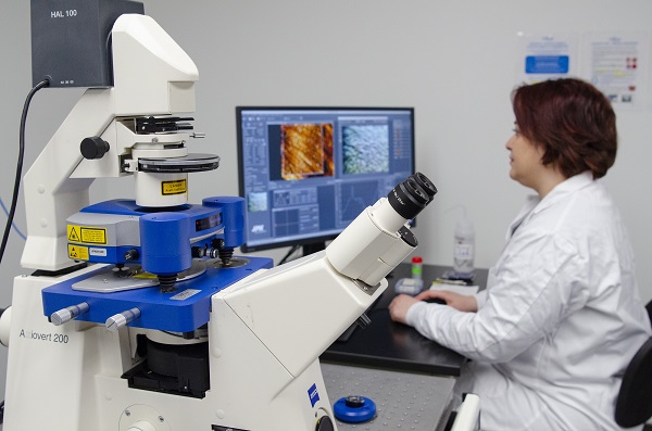
Atomic Force Microscopy
Atomic Force Microscopy (AFM) is a high-resolution technique that can visualize materials and cells at nano scale, and measure mechanical properties of a sample, such as viscoelasticity and adhesion strength.
Light microscopy
Light microscopy plays an important role in research, as it helps visualize fine details of an object. For samples that require crisp axial resolution, confocal microscopy is the recommended method, as it allows the three-dimensional reconstruction of the sample from images captured at different focal planes. CAMiLoD features several systems that can meet various lateral and axial resolution requirements, such as conventional, sub-diffraction and super-resolution confocal systems.
Explore our confocal microscopy systems.
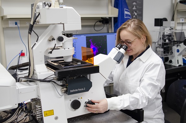
If there is no need for optical sectioning or a clear axial resolution, widefield microscopy equipped with a color CCD or monochrome CMOS cameras might be right choice to capture images quickly in brightfield and fluorescence.
Explore our widefield microscopy systems.
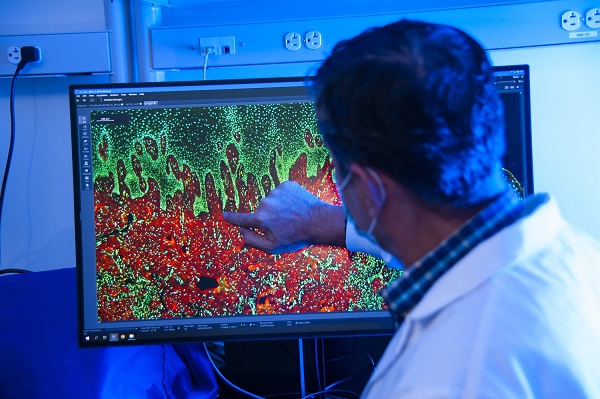
Histology
At the Histopathology research unit (HRU), experts study tissues and their structure. They can process, embed, section, and stain a variety of samples such as soft, hard, frozen and calcified tissues and implant materials.
HRU also provides immunostaining, histochemical staining, tissue microarrays and more. In 2022, this unit supported over 34 different projects.
More information on histopathology services.
“Histopathology is essential to the study of diseases. At CAMiLoD we provide a full spectrum of services, from tissue processing to staining and analysis, in one place, which can be quite convenient for our clients.”
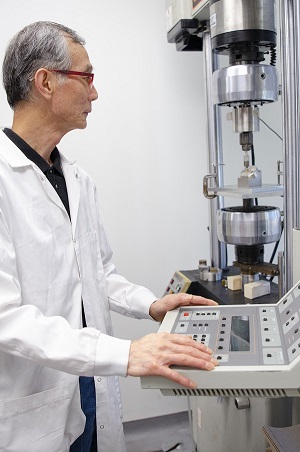
Mechanical Testing
CAMiLoD also offers fully serviced mechanical testing to assess fracture, fatigue, and microhardness properties of organic and synthetic materials based on the application of compression, tensile, or shearing force.
“Mechanical testing is an essential testing method that helps scientists with developing new materials.”
“For example, when developing new dental implants, researchers would conduct a fatigue test to determine how much masticatory force (force created during the act of chewing) would be required to damage an implant material.” says Jian Wang, manager of the Mechanical Testing and Scanning Electron Microscopy Units.
More information on Mechanical Testing services.
Scanning Electron Microscopy
Jian is also responsible for scanning electron microscopy (SEM) at CAMiLoD, where the Hitachi FlexSEM 1000 Scanning Electron Microscope is available for use.
“Going back to the dental implants example, a researcher might want to use mechanical testing first to break the implant material and/or hard tissue, and then use SEM to analyze the fracture in order to develop better biomaterials.”
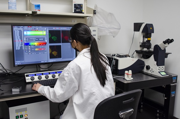
Slide scanning and image analysis
CAMiLoD also provides fully serviced and automated digitization of whole slide preparations. Users can submit the online requisition and leave routine histological, immunohistochemical, immunocytochemical, or immunofluorescence preparations to be scanned with a 20x or 40x objective. The scanned images can then be accessed on the dedicated CAMiLoD server for easy and efficient download from anywhere.
More information on slide scanning.
The team can also provide you with expert advice on ways and methods to better illustrate your science. They offer access to microscopy analysis programs like Volocity, Zen, Imaris (soon to come) and more.
More information on image analysis.
Interested in collaborating?
“The first step is to arrange a consultation with us, which is offered free of charge."
With so many different services available, from microscopy, to histology, to mechanical testing, it can be overwhelming to select the best methods and equipment for your research purposes.
“Choosing the optimum service or instrument depends on your research question. The first step is to arrange a consultation with us, which is offered free of charge. This will help us guide you on the best approach for your needs.” says Dhaarmini Rajshankar, Manager of CAMiLoD.
To learn more about CAMiLoD, visit CAMiLoD website. To book a consultation, please email camilod@utoronto.ca.
Written by Nina Ambros
Photos by Jeff Comber