
Unlocking the secrets of aging skin: Quantitative nanohistology
Latest research from professor Laurent Bozec and international collaborators, including five PhD candidates, demonstrated that by looking at variations in collagen properties in skin samples at the nanoscale, an individual’s biological age can be determined. Funded by the L’Oreal Group, the study focused on the quantitative analysis of aging dermal collagen using Atomic Force Microscopy (AFM). In collaboration between University College London, L’Oréal Research and Innovation, Lancaster University , Pontificia Universidad Catolica de Chile, U of T researchers explored the biophysical properties of dermal collagen at the nanoscale and differentiated between collagen from different age groups and anatomical sites. The study was published in the Frontiers in Aging journal.
The skin is the body's largest organ and plays a crucial role in protecting the body from environmental stressors. However, as we age, the skin undergoes complex changes that can affect its function, appearance, and overall health. “These changes are influenced by both intrinsic (chronological) and extrinsic (environmental) factors that can damage the skin's cells and extracellular matrix." says Bozec.
"Through this work we set out to understand the complex nature of connective tissue, such as skin, as a function of aging.”
The research team used AFM, a high-resolution microscopy technique, to examine the collagen network in skin samples from 30 female donors. They obtained high-resolution images of collagen fibrils and segmented them into smaller images for analysis. The collagen fibrils were classified based on four pre-defined empirical collagen structural biomarkers, which included gap formation, undefined collagen structure, and the presence or absence of dense collagen fibrillar networks with D-banding.
To complement the structural analysis, nanoindentation was performed on individual collagen fibrils from each section, resulting in a large dataset of indentation curves. Principal Component Analysis (PCA) was applied to reduce the complexity of the data and identify patterns.
“In this study, we proved that we could generate a large dataset using a localized probe imaging technique. This is an essential step for this kind of microscopy to support adjunct-diagnostic approaches.” says Bozec.
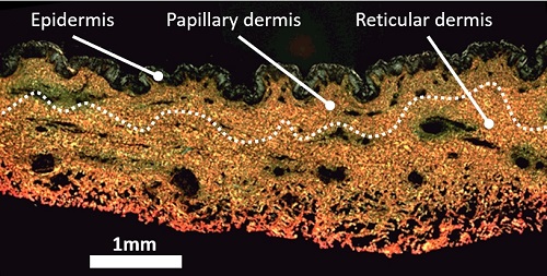
The prevalence of the empirical collagen structural biomarkers was compared between the papillary and reticular dermis of each section, allowing differentiation between donors based on their age or anatomical site.
"We are on course to create a new field of histology: quantitative nanohistology, to explore connective tissues’ structural and functional properties at the sub-micron level."
The study also included a case of abnormal biological aging, which validated the markers and nanohistology approach and highlighted the differences between chronological and biological aging in dermal collagen phenotyping.
“No established standards yet exist for collagen morphological variations at the nanoscale. This method of analysis would help in assessing chronic and pathological conditions that affect collagen structure and function.” says Bozec.
The study’s findings suggest that quantitative nanohistology can be a valuable tool for understanding connective tissue aging, differentiating between chronological and biological aging, and assessing collagen-related conditions.
Learn more about research at the Faculty of Dentistry.
Written by Nina Ambros
Photo credit: Jeff Comber IITS