Welcome to CAMiLoD
Faculty of Dentistry opens its door to research community with new, state-of-the-art imaging facilities
This is CAMiLoD: the collaborative advanced microscopy labs of Dentistry —and while it doesn’t have horses, knights or kings, this new state-of-the-art imaging facility could give rise to new legends of the research world.
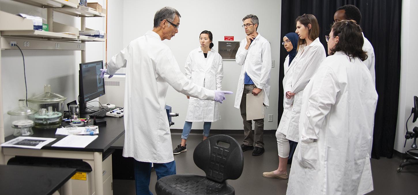
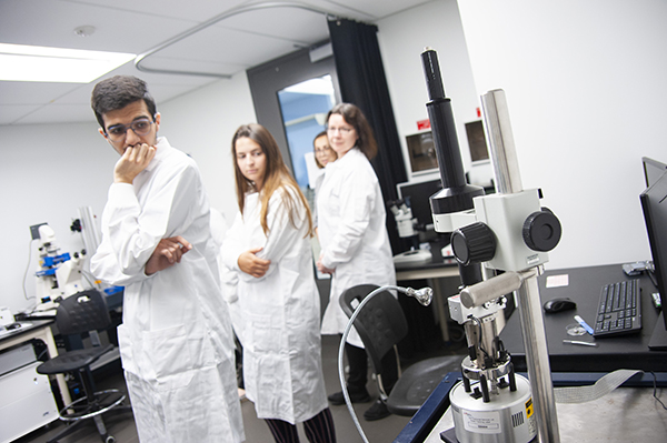
This week, with a full day symposium and workshop, the Faculty of Dentistry officially opened the doors of its new imaging facility to members of the research community.
“We wanted to collectively pool our equipment and make it useable for the entire community,” says university professor Boris Hinz, who spearheaded the project. Housed in Dentistry’s newly renovated research laboratories, CAMiLoD is where investigators of all stripes can rent time on some of the world’s best microscopy equipment to further their research.
"We wanted to collectively pool our equipment and make it useable for the entire community."
And the microscopy catalogue reads like a scientist’s paradise: looking to get up close with living cells and see them contract? The microscopy experts at Dentistry might recommend using the specialized upright confocal microscopes with water-dipping lenses. To view living cells in multiple positions over time, you might try to various Epiflourescence microscopes with fully automated Motorized Stage and on-stage incubation. Or do 3D imaging with living or fixed specimen with two scanning confocal microscope “workhorses,” the Leica SP8 and Zeiss LSM 800. But to see small surface structures, try the Hitachi Scanning FlexSem. Want to “see” forces at work? There’s the “Nanowizard 4” Atomic Force Microscope combined with correlative optical imaging. CAMiLoD even boasts a “Ferrari” of imaging products: the Zeiss LSM 880 Airyscan, allowing users to see their samples in 4D at high speed and sub-diffraction resolution.
Hinz hopes the labs will offer users more than imaging, though. Embedded within their existing services — light, force, and electron microscopy, as well as histology and image data analysis —CAMiLoD hopes to offer those working with hard-to-transport samples the option of using the shared cell culture and flow cytometry labs. To accommodate this service, CAMiLoD has also hired an imaging analyst, Joao Firmino, who can assist users of the facility with image post-processing and quantitative analysis.
“Pretty pictures are great but not enough,” says Firmino. “You need objective, quantitative data to get into the high-impact journals. We are here to help with this.”
“We hope this fully integrated approach —offering both cell culture and imaging —will be an asset to our research partners and students everywhere,” says Hinz. “Our team, which is coordinated by CAMiLoD manager Dhaarmini Rajshankar, has the technological and biological expertise to address your scientific problems,” he says.
“CAMiLoD is a creation that truly aligns with, and furthers, our Faculty’s vision to improve health by advancing dentistry through inspired leadership, innovation, and excellence in education, research and practice,” said dean Daniel Haas in a speech to kick off the symposium.
Thanking Hinz for his leadership, Haas added, “With CAMiLoD, faculty, students, and industry partners can come to the Faculty from all over the world to further their research, laying the ground for scientific and clinical collaborations, innovations and breakthroughs.
Interested in visiting CAMiLoD? Visit the website: https://www.camilod.ca
Images: registrants tour CAMiLoD, courtesy Jeff Comber